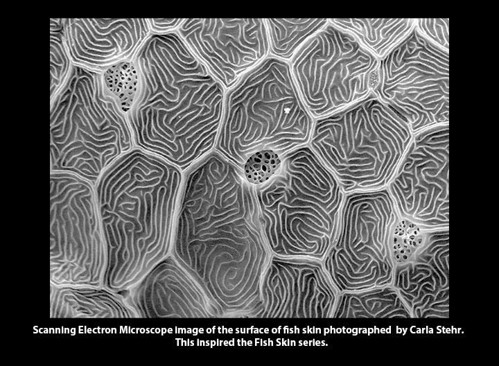
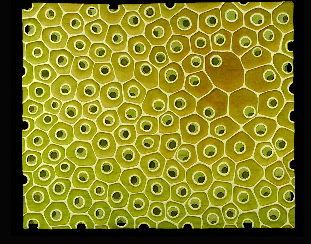
Fish skin is incredibly beautiful when seen with a scanning electron microscope. The surface of each skin cell has microridges reminiscent of fingerprint patterns. Specialized cells that produce mucus occur between the skin cells. They produce the "slime" that ordinarily covers the surface of the fish.

Below is the scanning electron microscope image of fish skin that inspired these pieces. This micrograph is from a newly hatched zebrafish. This fish is too young to have any slime in the mucus cells, providing an opportunity to see the surface of several skin cells and openings to three mucus cells.